Sunday, November 28, 2010

Haller cell

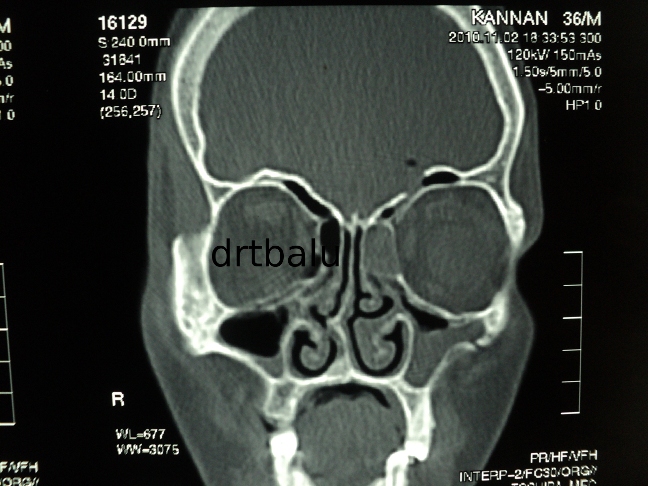
Synonyms: These are also known as Infra orbital recess cells.

Introduction:
These are pneumatized ethmoid air cells that project along the medial roof of the maxillary sinus and the most inferior portion of the lamina papyracea.
This air cell lies below the ethmoid bulla and lateral to the uncinate process.

Commonly these cells arise from anterior ethmoid air cells and are closely related to the infundibulum. Rarely these cells can arise from posterior
ethmoidal cells in which case it does not compromise the infundibulum.

Infections involving these cells can compromise and narrow the infundibulum causing obstruction to the drainage of maxillary sinus ostium.  It has been suggested that infections involving these cells could be a factor in recurrent maxillary sinusitis.


Thursday, November 25, 2010

Monday, November 22, 2010

CSF rhinorrhoea repair using bicoronal approach

Clinical details:

30 years old male patient came with c/o watery discharge from left nasal cavity - 3 years duration
H/O Road traffic accident 3 years back following which watery discharge started.
H/O meningitis 2 years back for which he was treated.
H/O Head ache on and off ++.

On examination:

Watery discharge was seen flowing out of left nasal cavity. It was more pronounced when the patient bent down.

Imaging:

CT paranasal sinus showed fracture involving the posterior table of frontal sinus with evidence of pneumatocele in the left
frontal lobe.


Management:

Since the fracture was present laterally in the posterior table of frontal sinus external approach was preferred.
Bicoronal approach was used to expose the anterior table of frontal sinus.
The incision begins in the preauricular crease at the level of tragus and traverses the scalp 3-4 cms behind the hair line.
The incision is carried down through the galea to the loose alveolar plane above the calvarial periosteum and the deep temporal
fascia and the flap is elevated anteriorly until approximately 2cms above the supra orbital rims. At this point the periosteum is
incised and the dissection is continued in the subperiosteal plane. The supraorbital neurovascular bundles are protected.
Laterally the dissection continues in the subgaleal plane up to 1-2 cms above the zygomatic arch. The deep temporal fascia
is incised at this point and the dissection continues deep to the temporal pad of fat over the temporalis muscle and the investing
fascia. The deep temporal artery perforators should be protected if not this could lead to a hallowing in the temoporal area due
to wasting of temporalis muscle.
The anterior table of frontal sinus is exposed. A window is created over the anterior table of frontal sinus using a fissure burr.
The interior of frontal sinus was visualized and the leak was identified over the posterior table of left frontal sinus which was
sealed using tissue glue and abdominal fat.


window.JPG


Friday, November 19, 2010

Interesting case of fibroma sphenoid sinus

Clinical details:

18 years old boy came with c/o blocking sensation both nasal cavity - 1 year duration
Decreased sensation of smell - 6 months duration
He gave h/o repeated attacks of upper respiratory infection
He gave no h/o bleeding from nose
No h/o head ache

On examination:

Anterior rhinoscopy:
A pale mass could be seen occupying the posterior portion of right nasal cavity.
Post nasal examination:
The same mass could be seen occupying the nasopharynx occluding both choana.

CT scan nose and paranasal sinuses plain / contrast were taken.

It showed non enhancing mass arising from sphenoid sinus entering the post nasal space and the posterior portion of right nasal cavity.


Management:

This patient was taken up for endoscopic sinus surgery and the mass was removed completely in toto.
Histopathology report - fibroma
Fibroma involving sphenoid sinus and post nasal space are commonly vascular (angiofibroma). In this case the fibrous elements
predominated and hence the mass was not enhancing in contrast CT. There was no abnormal bleeding during surgery.


Saturday, November 06, 2010

The Tamilnadu Dr MGR Medical University MS (ENT) Basic sciences march 2009 question paper with solution

In my continuing effort to help out post graduate students of otolaryngology I have uploaded the MS (ENT) Basic sciences question paper with solutions of Tamilnadu Dr MGR Medical university.

Basic sciences post graduate question papers have always been dicey.  Unless you prepare diligently for the same you may encounter pit falls.  The whole question paper has been broken down into sub topics i.e. anatomy, physiology, pathology and biochemistry.  Since each of these sections will be evaluated by different examiners belonging to basic sciences department you need to concentrate equally on all these sections.

You can access the question paper with solutions from here.