Complaints:
48 years old male patient came with complaints of loss of vision left eye - 6 months duration.Persistent left nasal block - 4 years
Discharge from left nose - 4 years
Head ache more on the left side - 5 years (Deep boring in nature)
Past history:
No history of epistaxisNo history of loss of smell
On examination:
Anterior rhinoscopy was unremarkable.Post nasal examination normal.
Vision was absent in the left eye.
Imaging:

<>
CT scan plain axial cut paranasal sinuses show heterodense mass arising from left posterior ethmoid air cells extending up to the anterior face of sphenoid. The mass could be seen compressing the left optic nerve. Diagnosis (Fibrous dysplasia).
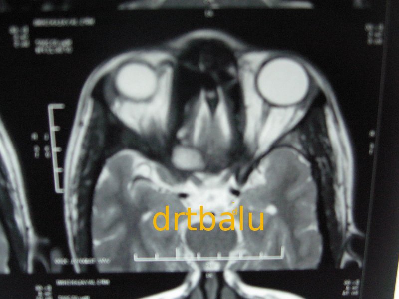
MRI showing mass arising from posterior ethmoidal air cells on the left side compressing the left optic nerve.
Management:
This patient was taken up for endoscopic surgery. The mass from the left side of the nasal cavitywas progressively drilled out under endoscopic vision.
Discussion:
The term fibrous dysplasia was first introduced by Litchtenstein in 1938. Usually fibrous dysplasias affect children commonlyin their teens (during the growth phase).
In any fibrous dysplasia involving the nasal cavity and paranasal sinuses the clinical presentation
is initially related to sinus obstruction, visual disturbance, facial asymmetry and nasal blockage.
In fibrous dysplasia the normal woven bone is replaced by isomorphous fibrous tissue and poorly
formed woven bone.
Classification:
Fibrous dysplasias have been classified into:Monostotic - Involves single bone
Polyostotic - Involves multiple bones
Albright's syndrome - Endocrine hyperfunction, Unilateral Café-au-lait spots, and polyostotic fibrous dysplasia
Among these types the monostotic fibrous dysplasia is more common accounting for 70% of all fibrous dysplasias.
Rarely fibrous dysplasia may become more aggressive and dedifferentiate causing (desmoplastic fibroma). Sometimes the tissue may undergo sarcomatous transformation.
Excision of these mases makes sense because of their propensity to undergo malignant transformation.
Risk factors for malignant transformation include:
1. Polyostotic form
2. Post radiation sequelae
3. Facial bone involvement
4. Albright's syndrome
No comments:
Post a Comment